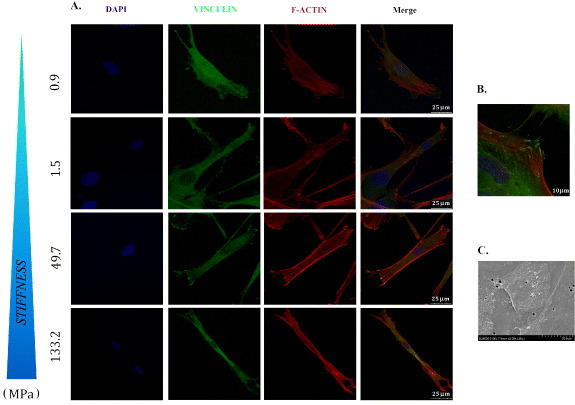
Figure 3.

Undifferentiated myoblasts functionally adhere to PCL films. C2C12 and MYB01 cells were cultured on PCL polymers with different stiffness values. Cell morphology and the formation of adhesion processes were detected by immunofluorescence staining after 24 h of culture. To visualize the cytoskeleton organization, F-actin was decorated by Phalloydin-TRITC (A, red). The expression of focal adhesion protein vinculin was detected independently from the layer (A, green). A detail of the focal adhesion processes is shown in (B). SEM analysis confirmed the undifferentiated morphology of adherent cells (C). Scale bars A: 25 μm; B: 10 μm; C: 20 μm.
