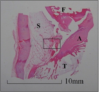
Figure 3.

Histology of knee joint (hematoxyline and eosin stain). (a) Sagittal view of knee joint 3 months after adhesive was applied. F: femur, A: anterior cruciate ligament, T: tibia, S: synovium. (b) Magnification of (a), synovium (hematoxyline and eosin stain). There was no inflammation in the synovium. (c) Meniscus of applying DST + HSA, 3 months after operation (hematoxyline and eosin). Arrows show the region where longitudinal tear was induced. (d) Magnification of (c) (hematoxyline and eosin stain). Crosslinking was seen in the ruptured meniscus, suggesting biological repair. (e) Meniscus of untreated control group 3 months after operation (hematoxyline and eosin stain). (f) Magnification of (e). Torn region is not repaired, there was no crosslinking.
