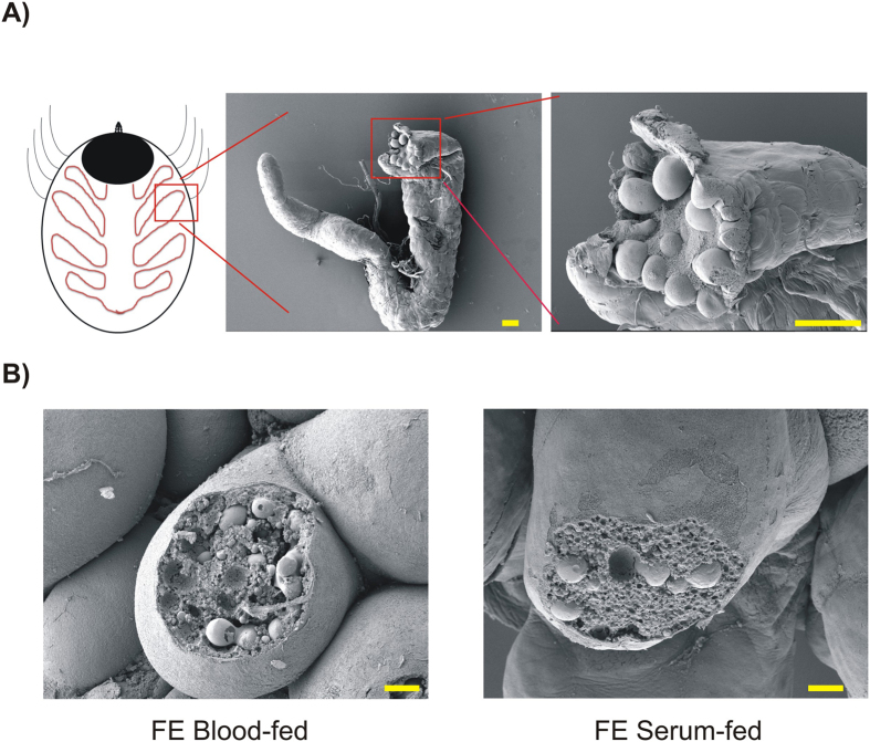
Figure 2. Scanning electron microscopy of tick gut caecum and digest cells.
(A) Illustration of tick gut caecum dissected from a partially-fed adult I. ricinus female. Such caeca were used for RNA-seq analyses. Scale bars indicate 100 μm. (B) Manually disrupted digest cells maturing along tick midgut epithelium from blood- (left) and serum-fed (right) fully engorged adult I. ricinus females. Note that digest cells from either tick contain both small and large digestive vesicles. Scale bars indicate 10 μm.

