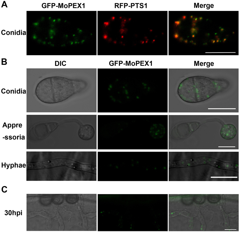
Figure 5. MoPex1 localizes to peroxisomes.
(A) Fluorescence of conidia from the co-transformant strain GDW2 expressing GFP-MoPEX1 and RFP-PTS1 was observed under a confocal microscopy. Bars = 10 μm. (B) MoPex1 expressed in the conidia, appressoria (incubated for 12 h) and vegetative hyphae. Bars = 10 μm. (C) The GFP-Mopex1 expression was observed in invasive hyphae at 30 hpi. Bars = 10 μm.

