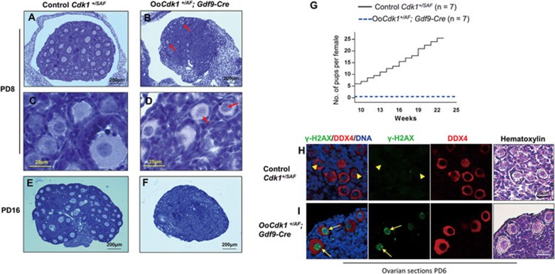
Figure 2.
Expression of Cdk1AF in dormant oocytes causes DNA damage and oocyte depletion in OoCdk1+/AF; Gdf9-Cre mice. (A-F) Hematoxylin-stained sections of mouse ovary at PD8 and PD16. By PD8, only a few follicles were left in OoCdk1+/AF; Gdf9-Cre ovaries (B arrows), and the oocytes of surviving primordial follicles remained arrested at GV stage (D, arrows). The follicular structures had mostly disappeared in the mutant ovaries by PD16 (F). As controls, normal ovaries of PD8 (A and C) and PD16 (E) Cdk1+/SAF mice containing healthy follicles are shown. The experiments were repeated more than three times each, and for each time and each age ovaries from one mouse of each genotype were used. (G) Comparison of the average cumulative number of pups per OoCdk1+/AF; Gdf9-Cre female (dotted blue line) and per Cdk1+/SAF female (solid black line), indicating that the OoCdk1+/AF; Gdf9-Cre females were infertile. The numbers of females used are shown as n. (H and I) Prominent γ-H2AX staining was observed in dormant oocytes enclosed in primordial follicles of PD6 OoCdk1+/AF; Gdf9-Cre ovaries (I, arrows) but not in the control oocytes (H, arrowheads). DDX4 was used to label the oocytes and the same sections were counterstained with hematoxylin to visualize ovarian histology. The experiments were repeated more than three times each.

