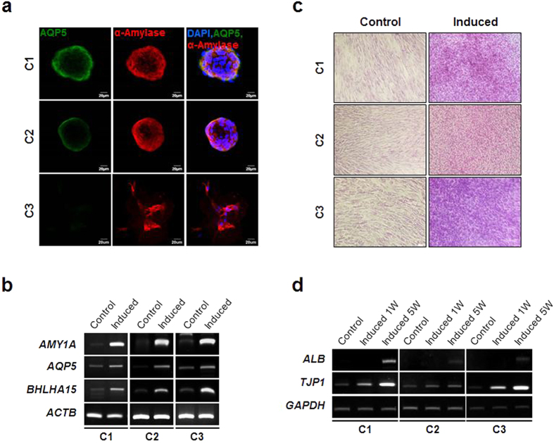
Figure 3. Epithelial differentiation potential.
(a) Each SG clone was allowed to form salisphere-like cell structure followed by salivary epithelial cell differentiation as described in the methods. Salivary epithelial differentiation was evaluated by immunofluorescence staining for AQP5 and α-amylase. Scale bars represent 20 μm. (b) Expression of salivary epithelial differentiation-associated molecular markers including AMY1A, AQP5, and BHLHA15 was examined by semi-quantitative RT-PCR. (c) To assess the multiple epithelial differentiation potential of the SG clones, each clonal population was subject to in vitro hepatogenic differentiation. The differentiation was evaluated by PAS staining. (d) Expression of hepatocyte differentiation markers including ALB and TJP1 was analyzed by RT-PCR.

