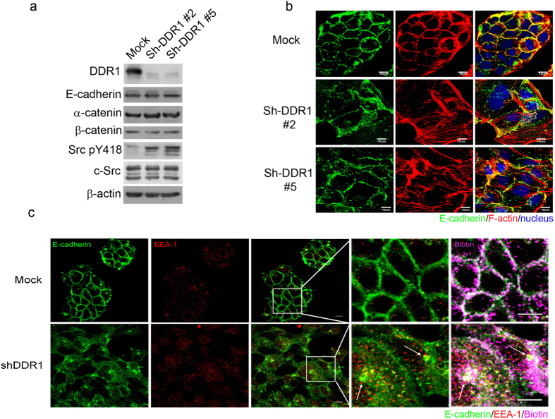
Figure 1. Knockdown of DDR1 decreases junctional localisation and increases endocytosis of E-cadherin.
(a) Control (Mock) and DDR1 knockdown (Sh-DDR1) MDCK 3B5 cells (Sh-DDR1#2, Sh-DDR1#5) were cultured on a culture dish for 24 h. The protein levels of DDR1, E-cadherin, α-catenin, β-catenin, Src pY418, and total Src were assessed using immunoblotting. (b) Localisation of E-cadherin (green) and F-actin (red) in Mock and Sh-DDR1 clones, and images were captured using a confocal microscope (Olympus FV-1000). Bar: 10 μm. (c) Mock (upper panels) and Sh-DDR1 (lower panels) cells were cultured on chamber slides for 24 h. The total cell membrane proteins were labelled using biotin, and endocytosis occurred at 37 °C for 30 min. Cells were then fixed and stained with E-cadherin (green) and EEA-1 (red). Biotins (pink) were labelled using Avidin-Alexa 594. Images were captured using a confocal microscope (Olympus FV-1000). Bar: 10 μm. Dashed rectangles were enlarged as indicated in the images on the right-hand side. Arrows indicate the colocalisation of E-cadherin, biotin, and EEA-1. Bars in the enlarged image: 5 μm.

