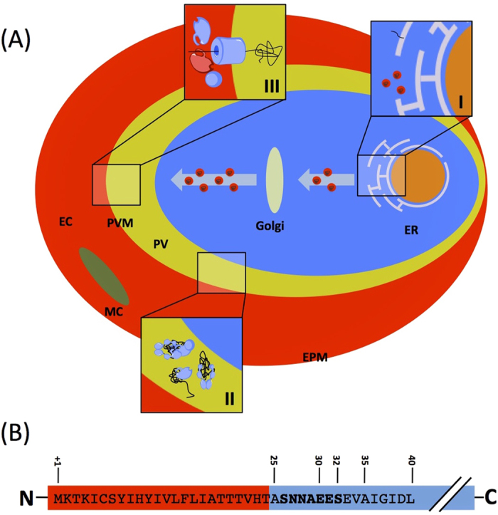
Figure 1.

(A) General model of protein export in the Plasmodium falciparum system. Proteins enter the secretory pathway at the ER, directed by an N-terminal secretory signal sequence or an internal hydrophobic segment (I). Proteins continue along the secretory pathway and are released into the lumen of the parasitophorous vacuole where they undergo unfolding, possibly facilitated by molecular chaperones (II). Following unfolding, these proteins are believed to then pass through a membrane bound translocon before emerging into the erythrocyte cytosol where they then refold and are carried to various localisations (III). ER, endoplasmic reticulum; PV, lumen of the parasitophorous vacuole; PVM, parasitophorous vacuolar membrane; EC, erythrocyte cytosol; MC, Maurer’s cleft; EPM, erythrocyte plasma membrane. (B) Structure of the N-terminal region of PfHsp70x. Red indicates predicted signal peptide. Numbers refer to amino acid from the N-terminus. The PfHsp70x export motif is shown in bold.
