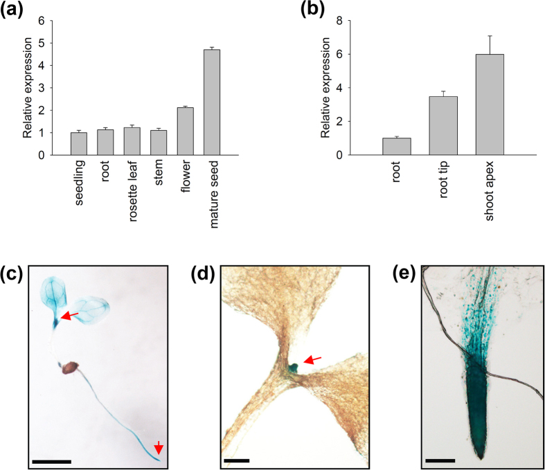
Figure 4. Expression pattern of AtMDN1.
(a) Relative expression analysis of AtMDN1 in the indicated tissues. Total RNA was isolated from 5-d-old seedlings, the stems of 40-d-old plants, the roots of 10-d-old plants, the rosette leaves of 20-d-old plants, the flowers of 60-d-old plants, and mature seeds (10 days after pollination). (b) Transcript abundance analysis of AtMDN1 in the root tip and shoot apex. Total RNA was isolated from the roots, root tips, and shoot apices of 10-d-old plants. (c) GUS staining analysis of AtMDN1 in a 5-d-old seedling. Bar = 5 mm. (d) GUS staining analysis of AtMDN1 in the shoot apex of a 5-d-old seedling. Bar = 1 mm. (e) GUS staining analysis of AtMDN1 in the root tip of a 5-d-old seedling. Bar = 100 μm.

