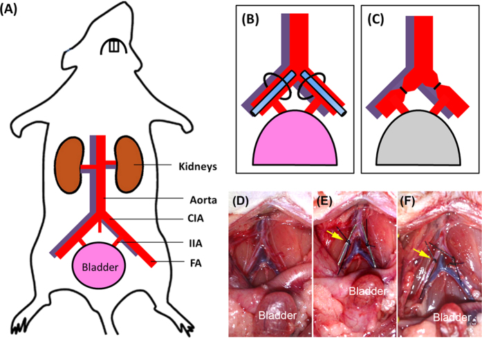
Figure 1. Illustration of animal model of bladder ischemia.
(A) Arterial supply of the bladder in rats. (B) The CIA was tied with a 3/0 silk ligature after PE-10 tubing was inserted through the FA. (C) Bladder blood flow decreased after partial occlusion by tubal ligation. (D–F) Experimental photos of the animal model before (D), during (E), and after (F) surgery. Yellow arrows indicate the partial arterial constriction. CIA, common iliac artery; IIA, internal iliac artery; FA, femoral artery.

