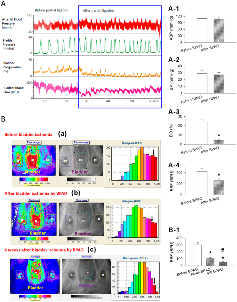
Figure 2.
(A) Representative recordings of changes in arterial blood pressure, bladder pressure, oxygenation, and blood flow in response to bladder ischemia. (A-1) Statistic data of the arterial blood pressure (ABP), bladder pressure (BP), bladder oxygenation (BO) and bladder blood flow (BBF) before and after bladder ischemia by partially common iliac artery ligation (BPAO). (B) Laser speckle contrast analysis of relative bladder microcirculation in response to bladder ischemia. Normal bladder blood flow is shown in flux image (left), photo image (middle), and perfusion histogram of ROI 2 (right). The distribution of bladder blood flow was significantly altered after the induction of ischemia, with a decrease in the high-flow area (red) accompanied by an increase in the low-flow area (blue). Three ROIs were established: ROI 1 (right femoral artery), ROI 2 (bladder body), and ROI 3 (left femoral artery). (B-1) Statistic data of the ROI 2 value displayed with blood perfusion unit in red color in the three groups of rats; before BPAO, acute BPAO and 4 weeks of BPAO. *p < 0.05, compared with sham treatment (before BPAO); #p < 0.05, compared with acute BPAO.

