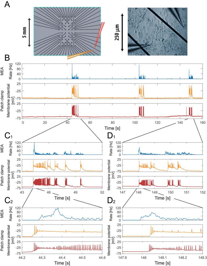
Figure 4. Extracellular multi-electrode array (MEA) recordings simultaneous with two distant neurons intracellular current-clamp recordings, indicating high correlation between cooperative burst of the network and single neuron membrane potential.
(A) Left: A zoom-in of the area covered by the extracellular electrodes, similar to Fig. 1A2. The orange and red electrodes represent two intracellular patch electrodes placed several millimeters out of the multi-electrode array. Right: A snapshot of part of the neuronal culture together with two neurons with two patch electrodes. (B) Similar to Fig. 2B. Upper panel: The rate activity of the multi-electrode array, over a period of 160 seconds, calculated using a convolution (see Methods). Middle and lower panels: The membrane potential of two current-clamped neurons placed several millimeters out of the multi-electrode array, (A), recorded simultaneously with the extracellular recordings shown in the upper panel. (C1) A zoom-in of the first burst shown in (B), for the network firing rate, recorded by the extracellular electrodes (top), and the voltage of the two patched neurons recorded by the intracellular electrodes (middle and bottom). (C2) An additional zoom-in of the 400 milliseconds at the beginning of the burst shown in (C1). (D1–2) Similar to (C1–2), for the last burst shown in (B). Results indicate a high degree of correlation between the extracellular recorded cooperative network activity and the two recorded membrane potentials.

