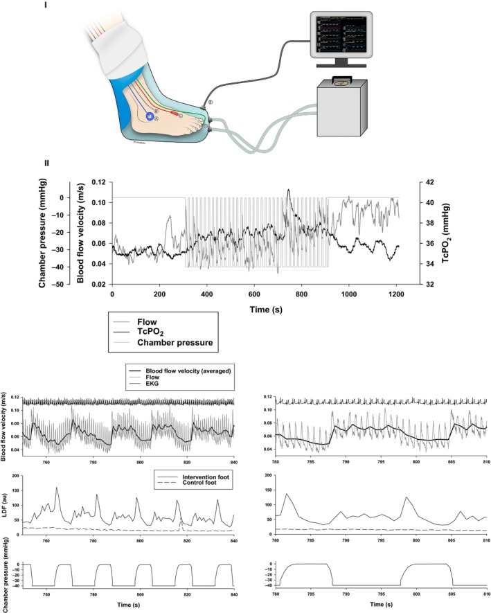
Figure 2.

I: Illustration of the custom‐made airtight vacuum chamber and the INP generator used by the four wound patients. The illustration shows how the probes were attached to the foot when measuring arterial blood flow velocity, laser Doppler flux (LDF), and transcutaneous oxygen pressure (TcPO2) in the foot of patient 1 during INP‐therapy. (A) TcPO 2 probe; (B) Skin temperature probe; (C) Ultrasound Doppler probe; (D) Laser Doppler flux probe; (E) The pressure transducer from the boot interfaced with the computer. Illustration: Øystein H. Horgmo, University of Oslo. II: Measures of acute hemodynamics in patient 1 after 8 weeks of INP. The upper large panel is TcPO 2 and arterial blood flow in the dorsal pedis artery (flow velocity) during the whole 20‐min sampling period: 5 min atmospheric pressure, 10 min INP and 5 min atmospheric pressure. Lower six panels: Beat‐to‐beat measures during application of intermittent negative pressure (INP) in patient 1 zoomed in from 750 to 840 sec (left) and from 780 to 810 sec (right). The panels show blood flow velocity and pulp skin flow response in the patient's right foot during application of INP. Upper panels: Blood flow velocity (ultrasound Doppler – thin lines), averaged within heartbeats (thick lines). Middle panels: Laser Doppler flux. Lower panels: Chamber pressure during INP.
