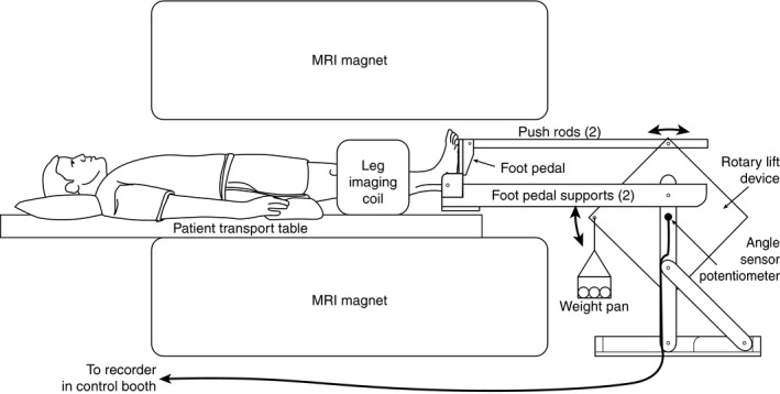
Figure 1.

Magnetic resonance imaging (MRI) compatible leg exercise device. The subjects were positioned supine with the largest part of the lower leg in the coil. Sand bags were added to the weight pan to increase the workload. A personal computer in the control room was connected via an MRI compatible cord to monitor the range of motion. This same device was also used for ultrasound studies (Protocols 1 and 3).
