
Fig. 4. Imaging through thick biological tissues.
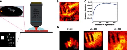
(A) Schematic of the experimental configuration. A positive resolution target USAF 1951 is placed in the focal plane of an immersion MO, with an 800-μm-thick layer of rat intestine on top of it. The region of interest is surrounded by a green square in the bottom inset. DIC, differential interference contrast; WD, working distance. (B) En face OCT image of the resolution target. (C) SD of the smart-OCT image as a function of the number M of eigenstates of Rs considered to build the image. (D to F) Smart-OCT images of the resolution target obtained from the 20, 250, and 500 first eigenstates of RS.