
Figure 1.
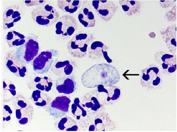
A cytospin of fixed CSF showing a N. fowleri trophozoite (arrow) stained with Giemsa-Wright amidst polymorphonuclear leukocytes and a few lymphocytes. Within the trophozoite, the nucleus and nucleolus can be seen. Magnification ×1000 [image is from CDC and can found at http://www.cdc.gov/parasites/naegleria/naegleria-fowleri-images.html]