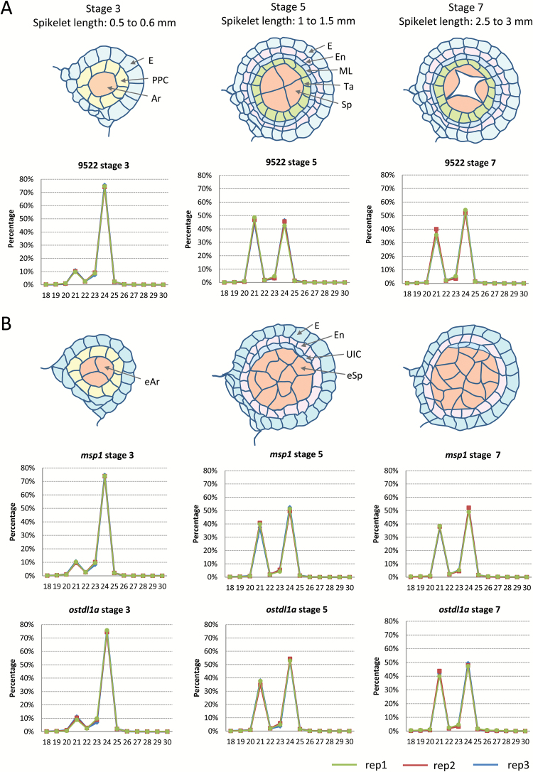
Fig. 1.
Small RNA size distribution in different developmental stages and backgrounds of rice spikelets. (A) Schematic representation of rice anther structures in different stages of spiklets of wild-type rice. Each layer of cells is indicated by an arrow: epidermis (E); primary parietal cell (PPC); archesporial cell (Ar); endothecium (En); middle layer (ML); tapetum (Ta); sporogenous cell (Sp). Small RNA size distributions in the different stages of wild-type rice spikelets are shown below. (B) Schematic representation of rice anther structures in different stages of spiklets of the msp1 and ostdl1a mutants. Excessive archesporial cell (eAr); unknown identity cell (UIC); excessive sporogenous cell (eSp). Small RNA size distributions in the different stages of the mutant spikelets are shown below.

