Fig. 1.
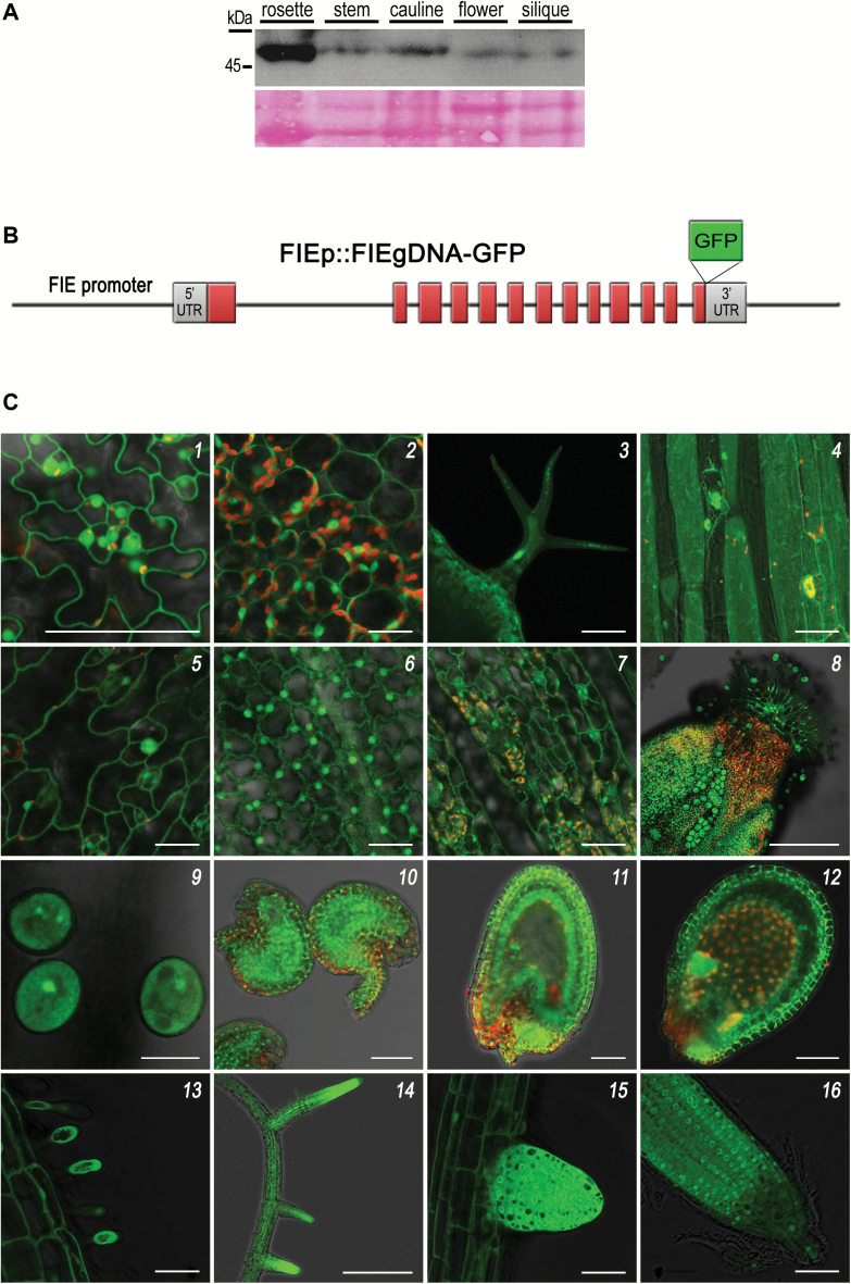
FIE protein accumulates in all Arabidopsis tissues and organs. (A) Detection of endogenous FIE protein in Arabidopsis tissues. A similar amount of nuclear chromatin protein extracts from rosette, stems, cauline, and inflorescence of 20-day-old wild-type plants and siliques from 6–10 days after pollination (DAP) were separated by SDS-PAGE and then immunodetected using αFIE antibodies. Protein size marker is indicated on the left. Ponceau staining was used to assess equal sample loading. (B) A scheme of the ProFIE:FIEgDNA-GFP transgene construct (not drawn to scale). Red boxes represent exons. (C) gFIE-GFP chimeric protein from line no. TH142-1 is observed in reproductive and vegetative tissues and organs. Each image was taken from a single focal plane, unless otherwise indicated. Each panel shows a merge of GFP epifluorescence (green) and the corresponding chloroplast autofluorescence (red) and/or a bright-field image. (1) Rosette leaf, abaxial epidermis; scale bar (SB): 50 μm. (2) Rosette leaf, mesophyll cells; SB: 25 μm. (3) Trichome on the surface of a rosette leaf, Z-stack overlay; SB: 50 μm. (4) Hypocotyl, Z-stack overlay; SB: 50 μm. (5) Cauline leaf, abaxial epidermis; SB: 25 μm. (6) Petal; SB: 25 μm. (7) Sepal; SB: 50 μm. (8) Carpel; SB: 250 μm. (9) Pollen; SB: 20 μm. (10) Unfertilized ovules; SB: 50 μm. (11) Developing seed containing a four cell-stage embryo; SB: 50 μm. (12) Developing seed containing a heart-stage embryo; SB: 100 μm. (13) Main root, root hairs; SB: 50 μm. (14) Main and lateral roots; SB: 500 μm. (15) Budding lateral root; SB: 50 μm. (16) Main root tip, quiescent center; SB: 50 μm.

