Fig. 3.
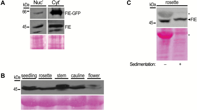
Endogenous FIE protein accumulates in the cytoplasm. (A) Nuclear (Nuc) and soluble cytoplasmic (Cyt) proteins were extracted from inflorescences of FIE-GFP plants, and analysed by SDS-PAGE followed by immunoblotting with αFIE and αGFP (Covance) antibodies. (B) Native cytoplasmic proteins were extracted from various tissues of wild-type Arabidopsis plants and equal amounts of samples were analysed by SDS-PAGE. The blot was probed with αFIE antibody. FIE was detected as a double band in all tissues, aside from rosette leaves. (C) Non-denaturated cytosolic protein extracts from rosette leaf tissue were sedimented by ultracentrifugation. Equal volumes from supernatant soluble fraction before and after (−/+) sedimentation were analysed by SDS-PAGE and probed with αFIE antibody. Absence of RuBisCO large chain protein band at ~53 kDa (marked with asterisk) following ultracentrifugation demonstrated successful sedimentation of large protein complexes. Ponceau staining in panels (A–C) was used to assess equal loading of samples.

