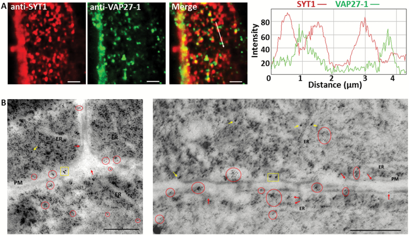
Fig. 3.
SYT1 and VAP27-1 accumulate on different regions of the ER–PM contact sites in Arabidopsis root cells. (A) Whole-mount immunocytochemistry of the root cells in wild-type Arabidopsis shows that SYT1 and VAP27-1 puncta are in close proxity on the cell cortex. The intensity profiles show that the peaks of SYT1 and VAP27-1 signals are shifted. Scale bars=2 µm. (B) Double immunogold labeling of SYT1 and VAP27-1 in Arabidopsis roots. The electron micrographs of ultrathin cryosections of the wild-type Arabidopsis root cells and immunogold labeling show that SYT1 (15 nm gold particles) and VAP27-1 (6 nm gold particles) are not localized on the same regions along the PM. The clusters of gold particles are highlighted as SYT1-clusters (yellow rectangles) and VAP27-1-clusters (red circles). SYT1 single labeling (yellow arrows) and VAP27-1 single labeling (red arrows) are indicated. Scale bars=500 nm.

