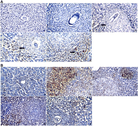
Fig. 4.

Mouse livers stained with TIMP-2 and Bax antibodies (a and b, respectively). a: Control group, with negative (−ve) TIMP-2 and Bax immunoreactivity in the hepatocytes and central vein. b: Vehicle control group, with negative (−ve) TIMP-2 immunoreactivity in the hepatocytes and strong positive (+ve) Bax immunoreactivity in hepatic tissues. c: PZQ (500 mg/kg bwt)-treated group, with mild TIMP-2 immunoreactivity and strong positive (+ve) Bax immunoreactivity in the hepatic tissues adjacent to the granuloma. d: CPE (300 mg/kg bwt)-treated group, showing positive (+ve) TIMP-2 immunoreactivity in the hepatic tissues surrounding the granuloma and mild Bax immunoreactivity in the hepatocytes adjacent to the fibrous granuloma. e: CPE (600 mg/kg bwt)-treated group, with positive (+ve) TIMP-2 immunoreactivity and weak Bax immunoreactivity in the hepatocytes adjacent to the fibrous granuloma (×400)
