
Fig. 2.
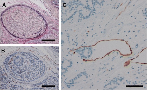
Representative images of venous and lymphatic invasion. Legend: a, b To determine the presence or absence of venous invasion, both EVG staining and immunohistochemistry for CD31 were performed. Venous invasion was confirmed, but no CD31-positive cells were identified. Such difficult cases were reviewed by more than two expert pathologists (EVG staining and immunohistochemistry for CD31; original magnification, ×200; scale bars represent 200 μm). c To determine the presence or absence of lymphatic invasion, immunohistochemistry for D2-40 was performed. Difficult cases were examined by more than two expert pathologists (Immunohistochemistry for D2-40; original magnification × 400; scale bar represents 100 μm)