Figure 17.

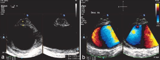
(a) Three-dimensional (zoom) image of the descending aortic short-axis view showing a 5 mm intramural hematoma in the wall of the descending aorta. (b) X-plane color Doppler imaging of the descending aorta showing the intramural hematoma

(a) Three-dimensional (zoom) image of the descending aortic short-axis view showing a 5 mm intramural hematoma in the wall of the descending aorta. (b) X-plane color Doppler imaging of the descending aorta showing the intramural hematoma