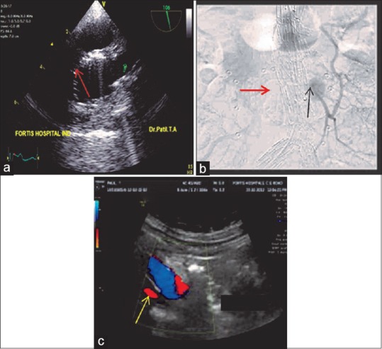
Figure 21.

(a) Upper esophageal aortic arch short-axis view demonstrating the (red arrow) endovascular stent in the aortic arch proximal to the origin of the left subclavian artery. (b) Aortography demonstrating a Type 2A endoleak (black arrow) into the aneurysmal sac from the inferior mesenteric artery. Red arrow is indicating the endovascular stent. (c) Transabdominal ultrasound color flow Doppler demonstrating a Type 4 (yellow arrow) endoleak
