Figure 5.

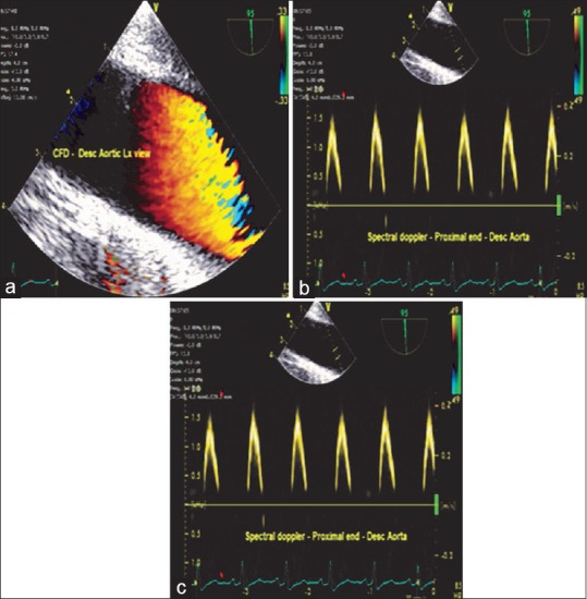
(a) Descending aortic long-axis view-color flow Doppler, (b) Spectral Doppler at the proximal and (c) distal end of the aorta depicted as above and below the baseline, respectively, indicating the flow toward and away from the transducer

(a) Descending aortic long-axis view-color flow Doppler, (b) Spectral Doppler at the proximal and (c) distal end of the aorta depicted as above and below the baseline, respectively, indicating the flow toward and away from the transducer