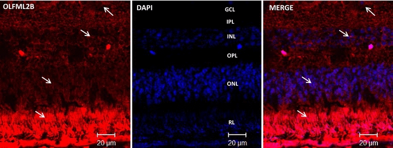
Fig. 4.

OLFML2A immunodetection in the retina of adult baboons. Confocal images of double stained retina sections to identify cells expressing OLFML2A (red 1st Ab: rabbit polyclonal anti-human OLFML2A 1:500; 2nd Ab: goat anti-rabbit IgG-Cy3® 1:4000), β-Tubulin (1st Ab: mouse monoclonal anti-mammal Tubulin 3 beta chain 1:250, 2nd Ab: goat anti mouse IgG FITC 1:250) and Glial Fibrillary Acid Protein (1st Ab: mouse monoclonal anti-GFAP 1:300, 2nd Ab: goat anti mouse IgG FITC 1:250) in baboon retina. Cells nuclei were labeled with DAPI (blue). GCL ganglion cell layer, IPL inner plexiform layer, INL inner nuclear layer, ONL outer nuclear layer, RL rod layer, PE pigmented epithelium
