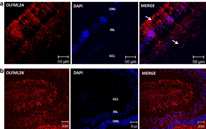
Fig. 6.

OLFML2A and OLFML2B immunoreactivity in human retina. a Confocal image of stained retina sections to identify cells expressing OLFML2A (red, 1st Ab: rabbit polyclonal anti-human OLFML2A 1:500; 2nd Ab: goat anti-rabbit IgG-Cy3® 1:4000). b Confocal image of stained retina seccions to identify cells expressing OLFML2B (red, 1st Ab: rabbit polyclonal anti-human OLFML2B 1:500; 2nd Ab: goat anti-rabbit IgG-Cy3® 1:4000). Cells nuclei were labeled with DAPI (blue). GCL ganglion cell layer, INL inner nuclear layer, ONL outer nuclear layer, PL photoreceptor layer
