Figure 5.
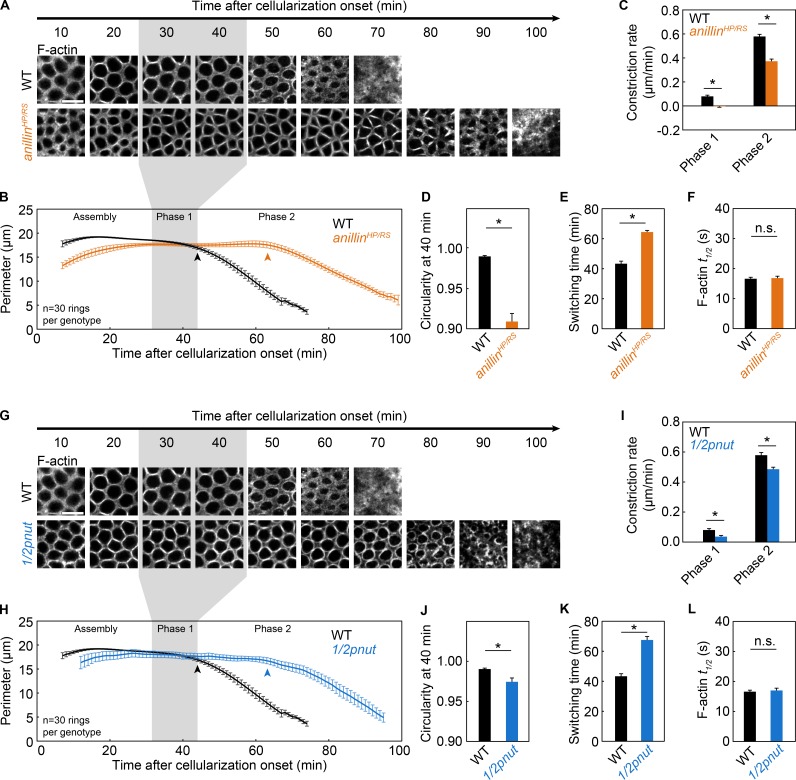
Anillin and Septin control timely switching to phase 2. (A–F) Live wild-type (WT; black) and anillinHP/RS (orange) embryos. (G–L) Live wild-type (WT; black) and 1/2pnut (blue) embryos. (A and G) Rings visualized with G-actinRed constricting over time (minutes). Bars, 10 µm. (B and H) Ring perimeter versus time. (C and I) Constriction rate per phase. (D and J) Circularity 40 min after cellularization onset. (E and K) Switching time from phase 1 to phase 2. (F and L) t1/2 for F-actin in photobleached rings (G-actinRed; F, n ≥ 35 rings from ≥14 embryos per genotype; L, n ≥ 16 rings from ≥6 embryos per genotype; mean ± SE; see Table S1). (A, B, G, and H) Gray shading highlights phase 1 in wild-type embryos. (B and H) Arrowheads indicate switching time. (A and G) Wild-type images are the same as in Figs. 3 G and 4 A. (B–E and H–K) n = 6 embryos per genotype and five rings were measured per embryo; mean ± SE. (C–F and I–L) *, P < 0.05; n.s., not significant.

