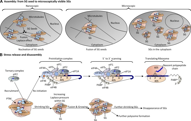
Figure 3.
Model of SG assembly and disassembly. (A) Once nanoscopic SG seeds are formed (Fig. 1 B), nearby seeds attract each other via weak electrostatic interactions, interact with neighboring SG seeds, and coalescence to form irregular microscopically visible SGs. Microscopically visible SGs can fuse to produce larger SGs. (B) After stress release, events that promote SG disassembly may include increase in concentrations of ternary complexes; phosphorylation of 4E-BP by mTOR, releasing the eIF4E block; and reactivation of eIF4A activities. These events might trigger the formation of translationally competent PICs to reinitiate translation at the surface of SGs. Because translating ribosomes displace mRNA-bound RNA-BPs, those complexes are then detached from SGs. As SGs shrink, the Laplace pressure increases to promote further fusion with adjacent SGs. Over time, fewer and larger SGs appear before they eventually disappear from the cytoplasm. Other SG proteins such as USP10 can potentiate disassembly by maintaining G3BP in a soluble conformation. PTM of ribosomal and SG proteins might also contribute to the disassembly.

