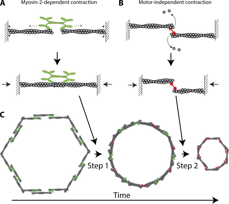
Figure 1.
Two-step model for actomyosin ring closure. (A) Model for Myo-2–dependent contraction. The motor heads of Myo-2 walk (green arrows) toward the barbed or plus ends of actin filaments (plus symbols). Because the forces on opposing heads are balanced, the actin filaments slide together, contracting the structure (black arrows). (B) Model for Myo-2–independent contraction. Two actin filaments held together by a cross-linking protein (red) depolymerize. Depolymerization brings the opposite ends of the actin filaments closer together, resulting in contraction (black arrows). (C) Back-to-back mechanism of ring closure. Step 1 represents the Myo-2–dependent step. Step 2 represents the Myo-2–independent step. Note that Myo-2 (green) and actin cross-linkers (red) are likely present during both steps, but are not included so as to illustrate the distinct steps.

