Fig. 1.

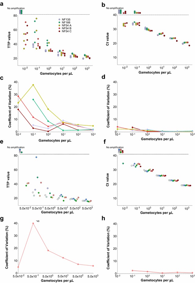
Intra (a–d) and inter (e–h) assay variation of QT-NASBA and qRT-PCR. In a–d, different colours represent different strains and cultures used for intra-assay variation assessment. In e–h, different experiments used in the inter-assay variation assessment are represented by different colours. Time to positivity (TTP) values for samples tested by QT-NASBA are presented in a and e and coefficients of variation (CVs), in c and g. Cycle of threshold (Ct) values for samples tested by qRT-PCR are presented in b and f; CVs are presented in d and h. In a, b, e and f, circles outside the y-axis range correspond to samples where Pfs25 mRNA was not detected. Of note, for experiments included in inter-assay variation analysis, different densities were used in gametocyte dilution series for QT-NASBA and qRT-PCR
