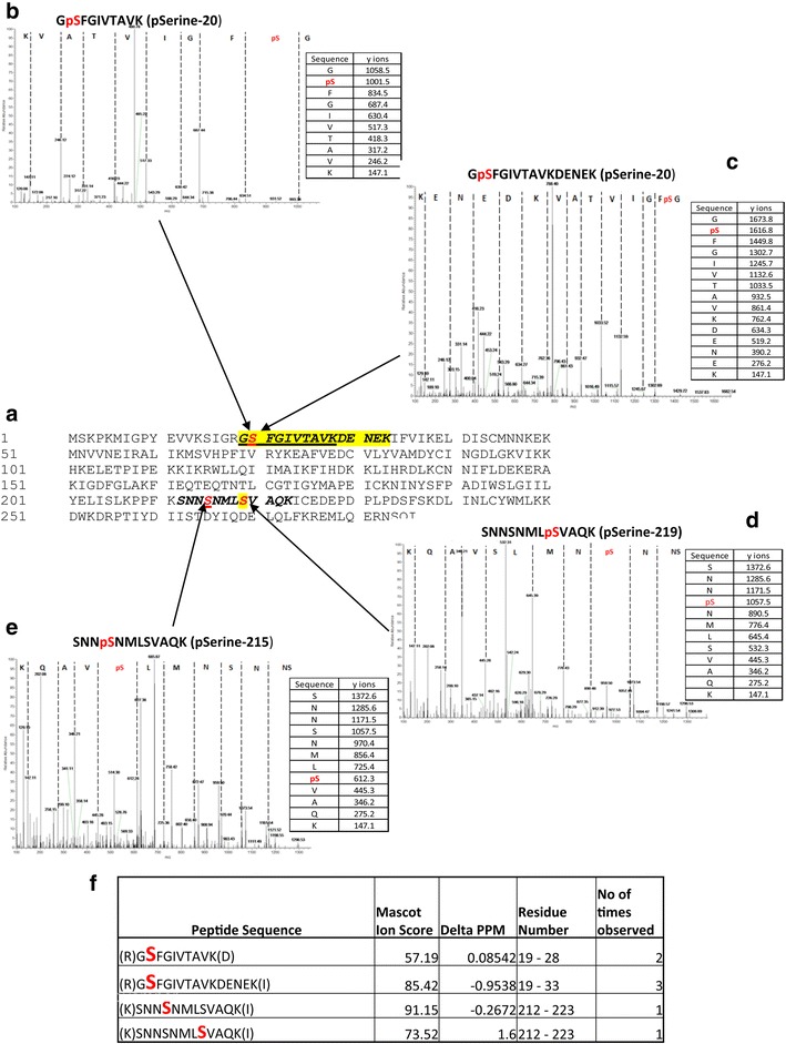
Fig. 7.

Autophosphorylation sites of Pfnek-2 as determined by mass spectroscopy. a Primary amino acid sequence of Pfnek-2. Sections in bold represent Pfnek-2 peptides observed in mass spectrometry experiments and in red are amino acids identified as being phosphorylated. b–e Representative mass spectra and associated fragmentation tables are shown that cover the three identified phosphorylated residues (Ser-20, Ser-215, and Ser-219). f List of the phospho-peptides identified in the overall data set
