
Figure 1.
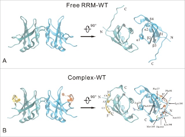
Cartoon representation of the free RRM domain (A) and the RNA-binding complex (B) systems. Two RRM domain (chain A and chain B) are colored cyan and blue, respectively. Chain P and chain Q of RNA are colored yellow and orange, respectively. The secondary structure elements are labeled on the free RRM domain. The important RNA-binding residues are highlighted in stick model, and the nucleotides are labeled on the RNA-binding complex.