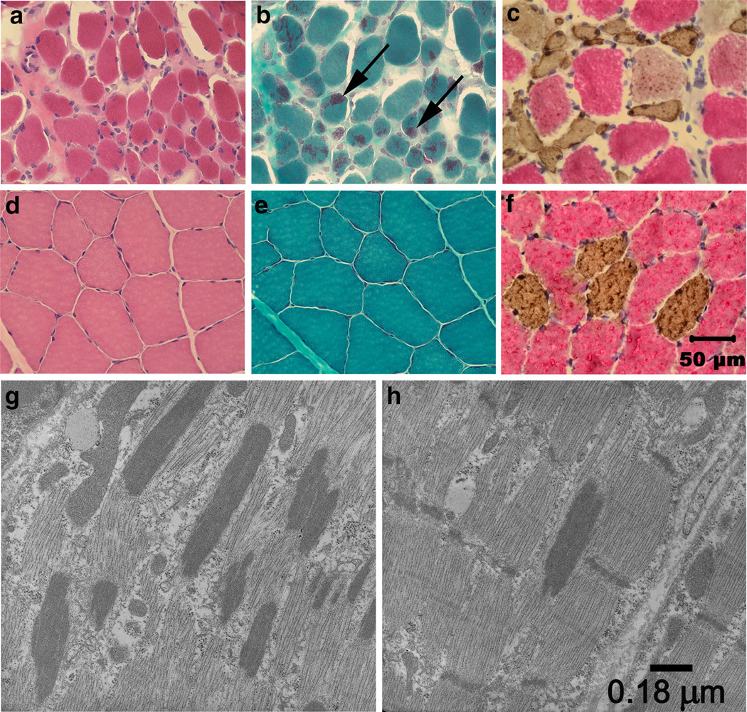
Fig. 1.
Histopathology establishes NM. Cryosections from the triceps muscle of an affected pup (a–c) and archived control triceps muscle (d–f) were stained with H&E (a, d), modified Gomori trichrome (b, e) and following incubation with monoclonal antibodies against type 1 and type 2 myosin heavy chains (c, f). Excessive variability in myofiber size and atrophy were observed in the affected muscle with the H&E stain (a) compared to control muscle (d). Numerous myofibers in the affected muscle contained rod bodies (b, arrows) not evident in control muscle (e). Both type 1 and type 2 fibers were atrophic (c) with fibers of both fiber types similar in size in control muscle (f). Bar 50 µm for images a–f. By electron microscopy, numerous electron dense rods were apparent along the long axis parallel to that of the muscle fiber (g). The rods were in structural continuity with Z disks (h), had the same electron density as the Z lines of adjacent sarcomeres, and had a similar lattice pattern of periodic cross striations. Bar 0.18 µm for images g and h

