Figure 3.
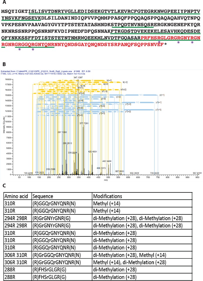
Identification of methylation sites in Scd6 by mass spectrometry. (A) Scd6 protein sequence showing arginine residues (marked with *) observed to be methylated in vivo by mass spectrometry analysis. Residues marked in red represent the RGG-motif (283–349). The green underlined residues represent the extent of protein coverage. (B) A representative mass spectrometry chart of GRGGQRGNYQNR peptide. (C) Table listing methylated peptides that were detected by mass spectrometry along with their methylation status.

