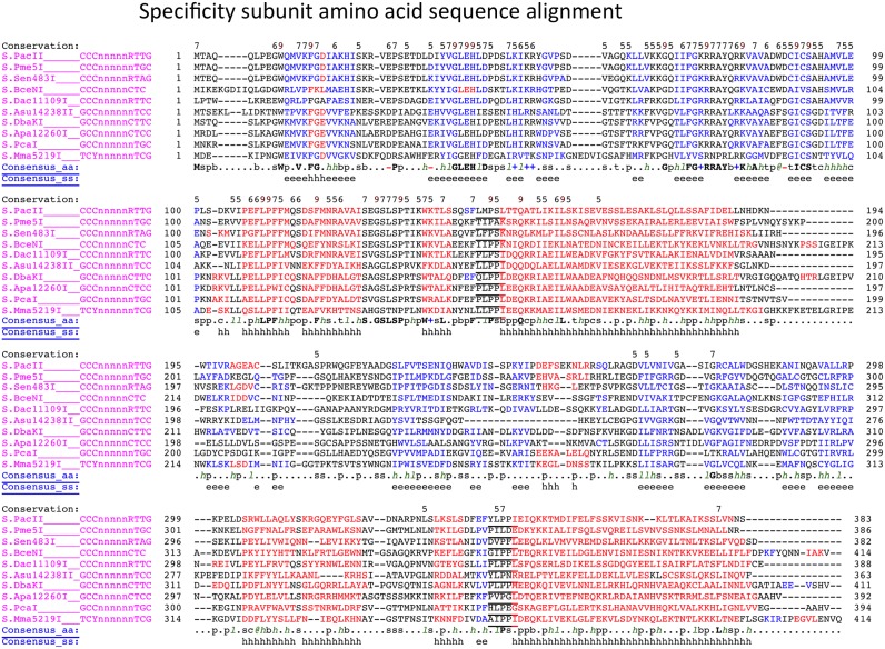
Figure 4.
Specificity subunit amino acid alignment. The conserved ‘PLPPL’ motifs connecting the globular TRDs into the two helical spacer sequences is underlined. Red indicates predicted helical secondary structure; blue indicates predicted beta strand secondary structure (from Promals alignment server).

