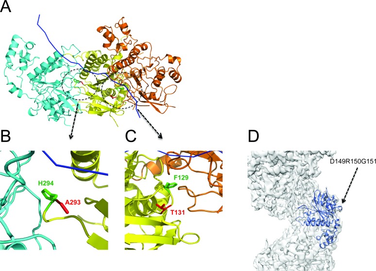
Figure 6.
Mapping of disease-associated point mutations onto the presynaptic filament structure. (A) For simplicity, three adjacent protomers shown here are coloured in yellow, cyan and orange, with ssDNA shown in blue ribbon. Residues A293 and T131 are coloured in red and residue H294 and F129 in green. (B) A close view at the residue A293. (C) A close view at the residue T131. (D) Mapping of mutations affecting Asp149, Arg150 and Gly151 on the EM map. These residues are situated on the outer surface of the RAD51 filament, distant from the protomer–protomer interface and the ATPase sites, and are unlikely to affect filament assembly.

