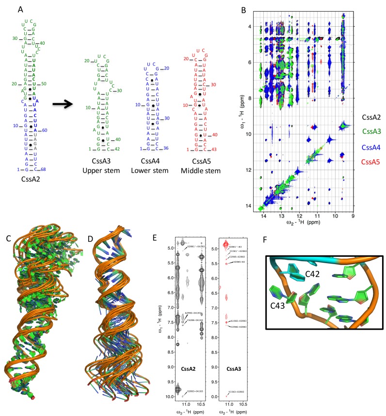
Figure 5.
NMR Assignment strategy and structural insight into CssA2 thermometer. (A) Three smaller constructs (CssA3, CssA4 and CssA5) were prepared to help structure determination of CssA2. Green and blue bold letters correspond to the 8 base sequence whereas gray colored bold nucleotides are the RBS. CssA3 corresponds to the upper stem of CssA2, CssA4 overlaps with the lower stem while CssA5 is representative of middle region of CssA. CssA5 was only used to refine the relative orientation of the upper and lower helices. (B) Overlay of the imino regions of 2D 1H-1H NOESY spectra recorded in 93% H2O and 7% 2H2O on Bruker Avance 800 MHz spectrometer at 7°C for CssA2, CssA3, CssA4 and CssA5. The black color corresponds to CssA2, green color is for CssA3, blue color for CssA4 and red color is used for CssA5. An excellent match is seen between each of the fragments (CssA3/4/5) and CssA2. (C) 1H-15N HSQC spectrum recorded in 95% H2O/5% 2H2O for CssA2 labeled with assigned peaks. The assignments were based on the spectra recorded for CssA2, CssA3, CssA4 and CssA5. Structural superposition of CssA2 RNA overlayed on (C) the lower helix (corresponding to CssA4) or (D) the upper helix (corresponding to CssA3). (E) Direct NMR evidence to support the U28*U41 base pair observed in NOESY spectra of CssA2 and CssA3, to demonstrate the similarities between the two constructs. (F) Orientation of C42 and C43 shown on the structure. The C43 base and ribose are projected toward the major groove, while the C42 ribose points inward, but its base has moved outside the helix, exposing both bases.

