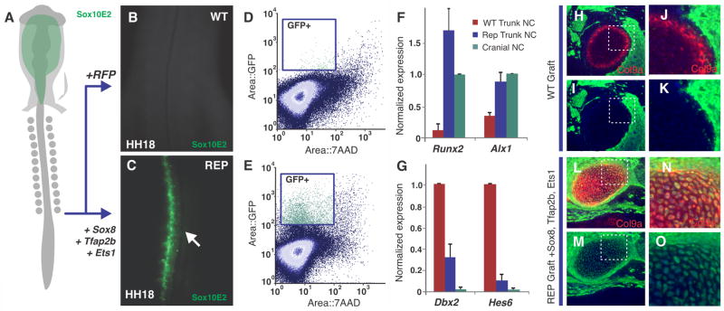
Figure 4. Reprogramming of neural crest axial identity and fate.
(A) Diagram of chick embryo electroporated with the Sox10E2 enhancer, expressed only in migrating cranial (Cr) neural crest (NC). (B) Transfection of trunk neural crest cells with control RFP expression vector shows no Sox10E2 expression. (C) Reprogramming of trunk neural crest cells with cranial specific regulators Sox8, Tfap2b and Ets1 results in robust expression of the cranial Sox10E2 enhancer in the trunk region (n=15/15). (D, E) Flow cytometry analysis of dissociated embryonic trunks shows a large number of Sox10E2+ trunk neural crest cells after reprogramming. (F, G) Reprogrammed (Rep) trunk neural crest display increased expression of the chondrocytic genes Runx2 and Alx1, while trunk genes Dbx2 and Hes6 are strongly downregulated. Error bars represent standard deviation. (H, I) Histological sections of E7 embryonic heads show that wild type (WT) trunk neural crest cells (GFP+; green) fail to form cartilage (Col9a+; red) after transplantation to the cranial region (n=0/5). (J, K) Inset of (H–I), showing absence of GFP+ chondrocytes. (L, M) Reprogrammed trunk neural crest cells (expressing Sox8, Tfap2b and Ets1) from GFP donor embryos transplanted to the head form ectopic cartilage nodules. (N, O) Inset of (L–M), showing chondrocytes derived from trunk neural crest cells (GFP+ and Col9a+).

