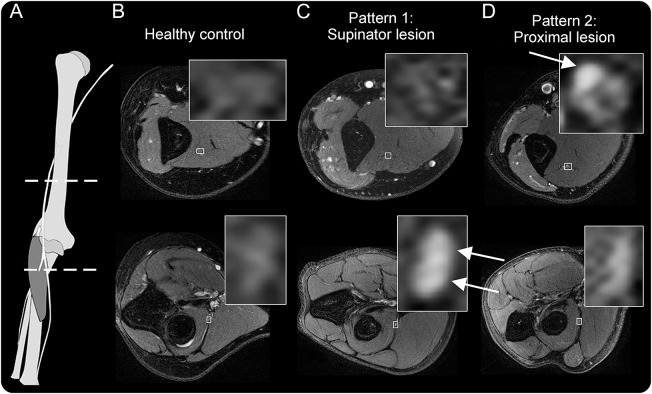
Figure 1. Two distinct lesion patterns in posterior interosseous nerve syndrome.
(A) Anatomical scheme of the radial nerve with dashed lines indicating the level of images. (B) Magnetic resonance neurography findings of a healthy control with normal radial nerve T2-weighted signal. (C, D) The first, expected pattern of findings for a patient presenting with posterior interosseous neuropathy syndrome: proximally at the upper arm level, a normal appearance and signal of the radial nerve, while after bifurcation and at entry into the supinator muscle, the nerve shows severely increased T2-weighted signal of the deep branch (arrow), in this case with little caliber increase. (D) A patient with the second lesion pattern: a proximal fascicular lesion, involving only a portion of the radial nerve trunk at the upper arm level (arrows).

