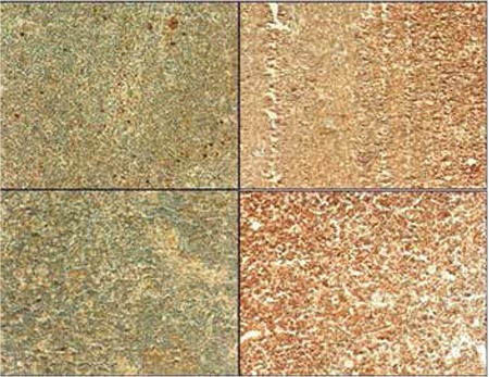
Figure 1. Histopathologic images of ROCK staining. Immunohistochemical staining for lymph node tissues with ROCK1 in control (a) and in mantle cell lymphoma patients (b), and ROCK2 staining in control (c) and in mantle cell lymphoma patients (d). Original magnification 200x.

