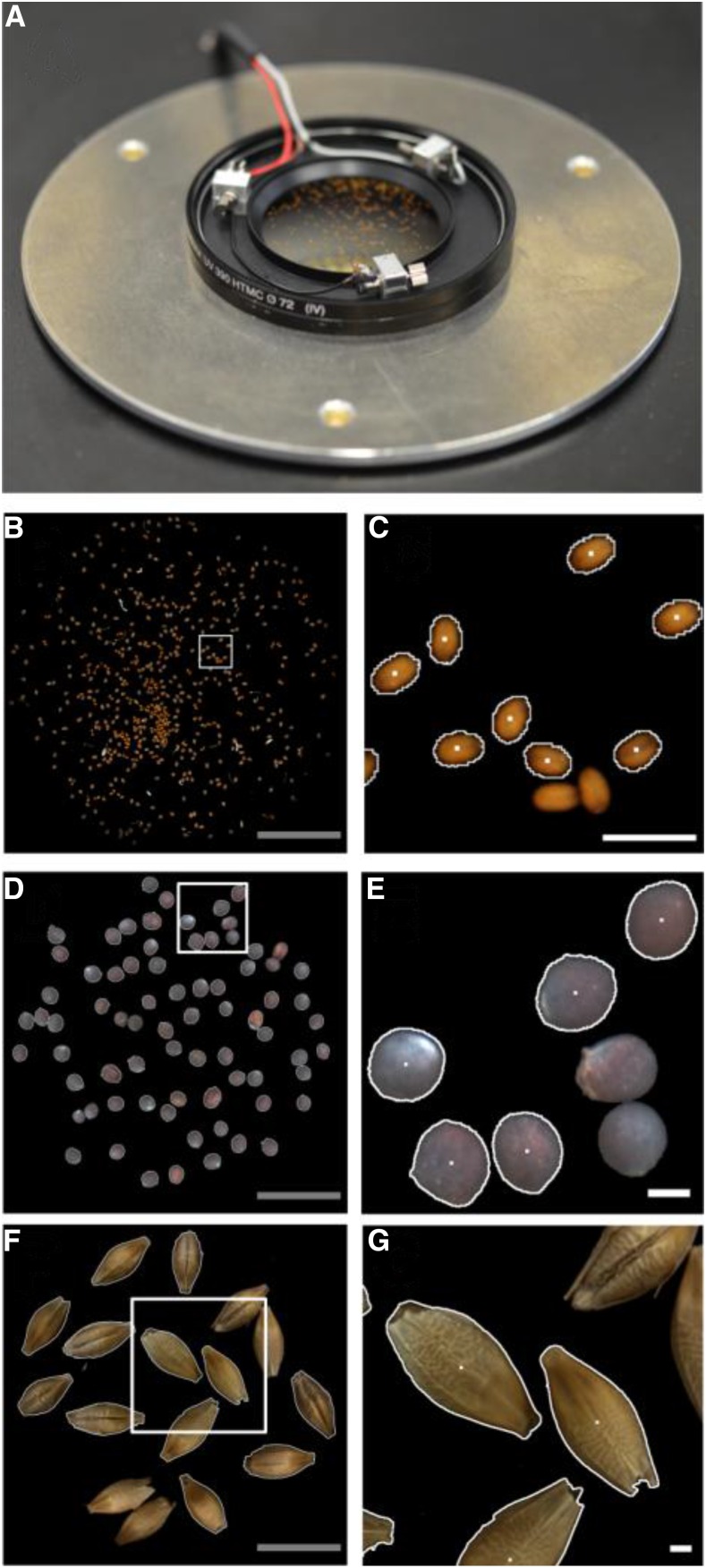
Figure 3.
2D imaging station. A, Module for 2D imaging with an optical glass filter on which seeds are dispersed and with three vibration motors to separate seeds lying close together. Below the glass filter, a light-emitting diode ring for illumination and a camera are mounted (shown in Supplemental Fig. S2). B to G, Seed images of Arabidopsis (B and C), rapeseed (D and E), and barley (F and G). B, D, and F, Total field of view with a diameter of 41 mm. C, E, and G, Enlarged areas (indicated by white squares in B, D, and F, respectively) where segmented seeds (indicated by white borders) and nonsegmented seeds (without white borders; these are skipped to ensure single-seed handling) are shown. White dots denote the center of gravity of the segmented seeds. Gray bars = 10 mm; white bars = 1 mm.

