Figure 4.
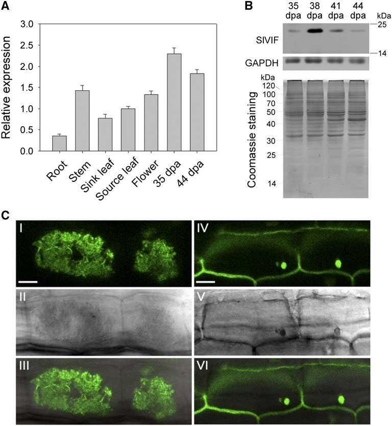
Characterization of tomato SlVIF. A, qRT-PCR analyses of SlVIF in vegetative and reproductive tomato organs. The ACTIN gene was used as an internal control. Values are the means of three biological replicates. Bars represent standard deviations. B, Western-blot analysis of SlVIF protein abundance during fruit ripening. Proteins were isolated from fruit at 35 DPA, 38 DPA, 41 DPA, and 44 DPA. An anti-glyceraldehyde-3-phosphate dehydrogenase immunoblot was used as a protein control. In addition, Coomassie Brilliant Blue staining was used as a loading control. C, Subcellular localization of GFP:SlVIF fusion protein. I, to III, Stable expression of the GFP:SlVIF fusion protein in tomato root cells, showing fluorescent signal in the vacuole. IV to VI, Stable expression of GFP alone in tomato root cells, showing fluorescent signals throughout the cell. I and IV show fluorescent images; II and V are the same images viewed using bright-field microscopy. III and VI are overlays of fluorescent and bright-field images. Bar = 10 μm.

