Figure 7.
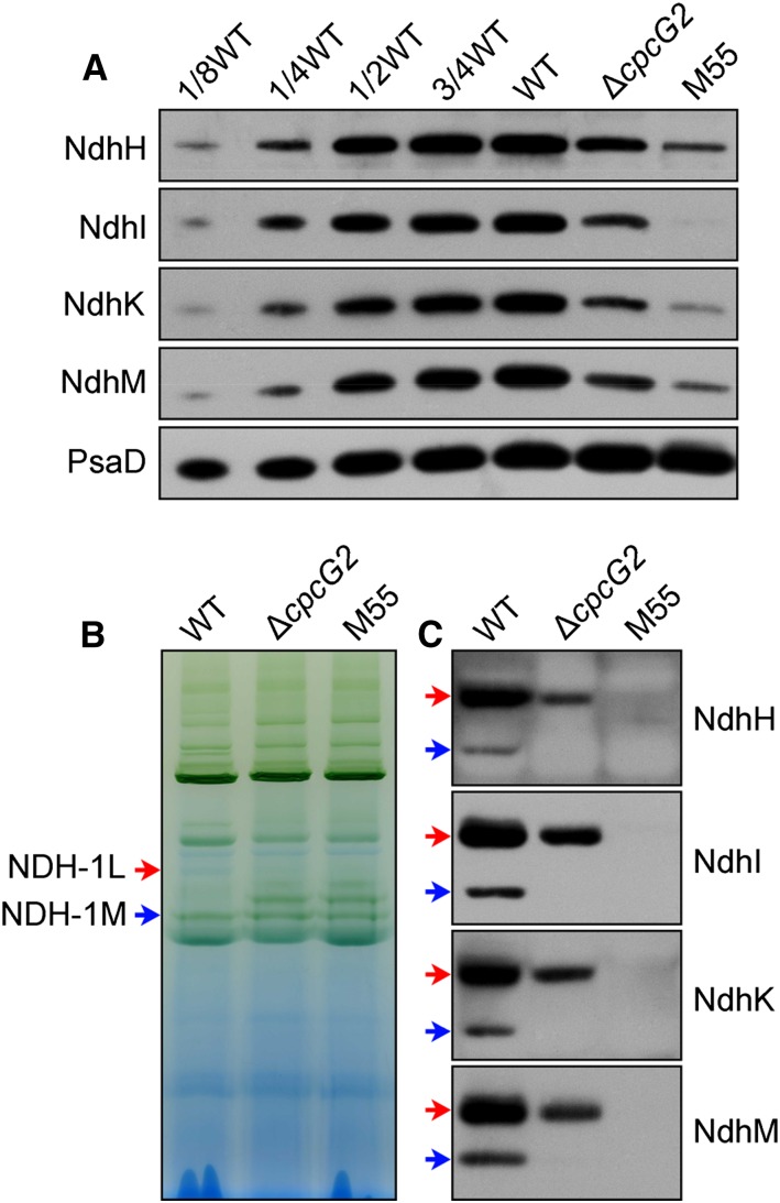
Accumulation and assembly of NDH-1 complexes. A, Immunodetection of Ndh subunits in the thylakoid membranes from the wild type (WT; including the indicated serial dilutions) and ∆cpcG2 and M55 mutants. Protein blotting was performed with antibodies against several hydrophilic Ndh subunits (NdhH, NdhI, NdhK, and NdhM). Lanes were loaded with thylakoid membrane proteins corresponding to 1 µg of Chl a (100%). PsaD was used as a loading control. B, BN-PAGE profiles of the wild type and mutants used in A. Thylakoid membrane extract corresponding to 9 µg of Chl a was loaded on each lane. Red and blue arrows indicate the positions of NDH-1L and NDH-1M complexes, respectively. C, Western-blot analysis of the BN gel in B with antibodies against specific Ndh subunits, showing a cross-reaction with the NDH-1L and NDH-1M complexes.

