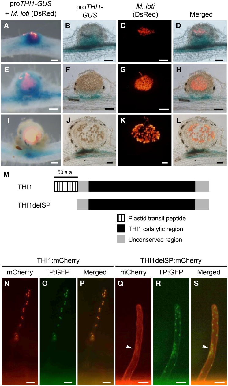
Figure 4.
Spatiotemporal expression of THI1 during RN symbiosis and subcellular localization of THI1. GUS staining is shown for transgenic hairy roots carrying the proTHI1-GUS construct inoculated with M. loti expressing DsRed. A, E, and I, Whole nodules. B to D, F to H, and J to L, Nodule sections of 100 μm. A to D, Nodule primordia. E to H, Young nodules. I to L, Mature nodules. Bars = 100 μm. M, Schematic diagram of the THI1 structure. The truncated protein THI1delSP has a deletion of the plastid transit peptide (50 amino acids [a.a.]). N to P, Coexpression of THI1:mCherry and GFP fused with a fragment encoding the plastid transit peptide of Arabidopsis Rubisco (TP:GFP) in L. japonicus hairy roots. Q to S, Coexpression of THI1delSP:mCherry and TP:GFP. Arrowheads indicate the nucleus. Bars = 20 μm.

