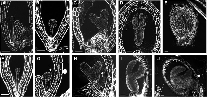
Figure 2.
Embryo development in tws1-1. A to J, Longitudinal sections of seeds at different stages of development imaged through the modified pseudo-Schiff propidium iodide imaging technique. Wild-type (A) and tws1-1 (F) seeds at the early globular stage of embryo development. Wild-type (B) and tws1-1 (G) seeds at the globular stage of embryo development. Wild-type (C) and tws1-1 (H) seeds at the heart stage of embryo development. Wild-type (D) and tws1-1 (I) seeds at the torpedo stage of embryo development. Wild-type (E) and tws1-1 (J) seeds at the mature stage of embryo development. Scale bars = 40 µm. Arrows point to sites where tws1-1 embryo epidermal cells fuse to the surrounding tissues.

