Figure 5.
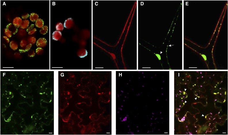
TWS1 is localized to the ER. A, Confocal image of a mesophyll-derived protoplast transformed with a construct carrying TWS1 fused to GFP (Pro-35S:TWS1:GFP). GFP signal is shown in green, chlorophyll signal in red. Scale bar = 5 µm. B, Confocal image of a mesophyll-derived protoplast transformed with a construct carrying VMA21 fused to CFP (Pro-35S:VMA21:CFP). CFP signal is shown in cyan, chlorophyll signal in red. Scale bar = 5 µm. C to E, Confocal images of stably transformed Arabidopsis plants carrying the construct Pro-35S:TWS1:GFP. FM4-64 signal (C), TWS1::GFP signal (D), and overlay of the FM4-64 and GFP channels shown in C and D (E). Scale bars = 20 µm. F to I, Confocal images of stably transformed tobacco mesophyll cells expressing RFP fused to an ER retention signal, infiltrated with the TWS1::GFP construct. F, TWS1::GFP signal; G, ER-localized RFP signal; H, chlorophyll autofluorescence; I, superimposition of images in F to H. Arrowheads point to a few of the positions where GFP and RFP colocalize. Scale bars = 10 µm.

