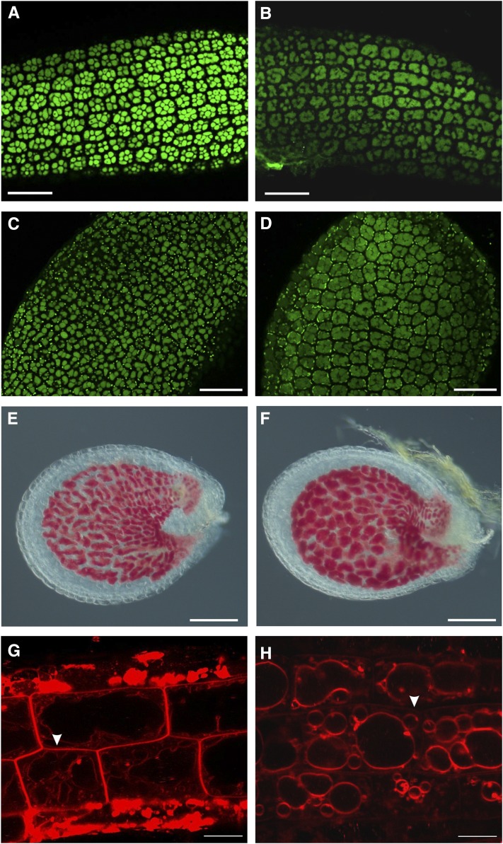
Figure 8.
Altered vacuole morphology in tws1. A to D, Confocal images of protein autofluorescence in embryo hypocotyls (A and B), and cotyledons (C and D). A and C, Wild-type embryos; B and D, tws1-1 embryos. Scale bars = 40 µm. E and F, Wild-type (E) and tws1-1 (F) vanillin-stained seed. Both seeds contain embryos at the heart stage. Scale bars = 100 µm. G and H, Confocal images of FM4-64-stained root epidermal cells of wild-type (G) and tws1-1 (H) seedlings. Scale bars = 10 µm.

