
Figure 3.
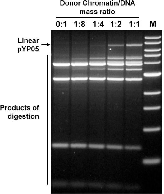
Characterization of chromatin templates using restriction enzyme sensitivity assay. Chromatin was assembled on supercoiled pYP05 plasmid and digested with an excess of restriction enzymes DraI and BglII. Then DNA was purified and analyzed in 1% agarose gel. Nucleosomes protect DNA from digestion with restriction enzymes. The restriction sites are localized beyond the NPSs (Fig. 1B); therefore the increase of donor-chromatin/DNA ratio results in progressively better protection of the template DNA from the enzymes, but this protection is still minimal even at highest ratio of donor chromatin to DNA (1:1), indicating that the majority of nucleosomes were formed predominantly on the desired NPSs. Only a small fraction (<5%) of all nucleosomes formed on the plasmid occupy the NPS-free regions of the pYP05 plasmid. M – 1-kb DNA ladder (New England Biolabs).