
Figure 4.
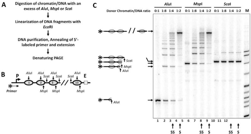
Analysis of chromatin templates using a restriction digestion sensitivity assay with primer extension. A. The experimental approach. B. Schematic diagram of the fragment of pYP05 plasmid used for in vitro chromatin reconstitution. Nucleosome positioning sequences (NPS) are shown by solid ovals. Restriction enzyme sites within the NPSs are shown by arrows. AluI and MspI sites are localized within NPS, ScaI sites - in the middle of NPS-separating spacer DNA. MspI and ScaI sites are missing from the first NPS and the spacer DNA respectively. C. Analysis of the products of primer extension by a denaturing PAGE. The increase in the level of chromatin assembly results in progressively better protection of the templates from AluI and MspI, but not from ScaI restriction enzyme. Possible products of digestion are shown on the right. S/SS: Saturated/sub-saturated chromatin samples.